
emDOCs.net Emergency Medicine (EM) Podcast
emDOCs.net Emergency Medicine (EM) Podcast
Episode 17: Sick Meningitis, POCUS for Pneumoperitoneum, and Treatment of CHS
Use Left/Right to seek, Home/End to jump to start or end. Hold shift to jump forward or backward.
Welcome to the emDOCs.net podcast with Brit Long, MD (@long_brit) and Manpreet Singh, MD (@MprizzleER)! Join us as we review our high-yield posts from our website emDOCs.net.
Today on the emDocs cast with Brit Long, MD (@long_brit) and Manpreet Singh, MD (@MprizzleER) we cover three posts: the sick meningitis patient, ultrasound for pneumoperitoneum, and treatment of cannabiniod hyperemesis syndrome.
To continue to make this a worthwhile podcast for you to listen to, we appreciate any feedback and comments you may have for us. Please let us know!
Subscribe to the podcast on one of the many platforms below:
emDocs Podcast Script:
Brit: Thanks for joining us on the emDocs.net podcast. I’m Brit Long, and I’m joined by Manny Singh. Today’s podcast covers three posts: the sick meningitis patient, US for diagnosis of pneumoperitoneum, and cannabinoid hyperemesis syndrome.
Manny: Our first post today published on Oct 7, 2019 is on managing the sick patient with meningitis.
We are trained how to evaluate and treat the patient with suspected meningitis, but the sick meningitis patient is challenging. Severe cases are associated with severe morbidity and mortality.
These patients will typically report at least 1 of the classic symptoms of meningitis (fever, neck stiffness, headache) and also have severe myalgias in nearly all reported cases. These patients will also have the classic petechial/purpuric rash on presentation only 50% of the time. On exam, these patients may also present with pallor and mottling of the skin up to 70% of the time, which should be regarded as an indication of severe systemic illness. The most deadly form of skin involvement secondary to meningococcal infection is purpura fulminans, an acquired, often fatal, coagulopathy characterized by extensive skin involvement which rapidly progresses to skin necrosis with disseminated intravascular coagulation (DIC).
These patients need resuscitation and may benefit from fresh frozen plasma (FFP) (10-20 ml/kg every 8 hours) and/or protein C concentrate (100-120 units/kg initial bolus). Protein C concentrate may help in these patients to help restore the balance of coagulation and anticoagulation factors which have become deranged in DIC. Two clinical trials have been conducted on a small population of patients with meningococcal meningitis, which have been associated with correction of coagulopathy without significant adverse events, as well as reducing the rate of amputation in these patients.
Brit:
If soft tissue/skin necrosis is found, consultation with general surgery is recommended as debridement, fasciotomy, and/or amputation may be indicated based on progression of illness14. In addition, the DIC and coagulopathy that occurs secondary to severe septicemia can also lead to thrombosis and hemorrhage of internal organs and exsanguination15. Patients may require aggressive volume expansion with crystalloid and blood products and intervention according to organ involvement such as intubation for respiratory failure from massive hemoptysis, surgical removal of infarcted internal organs, interventional radiology (IR) consultation for embolization, stress dose steroids for adrenal hemorrhage, or emergent hemodialysis for renal failure.
Another complication of meningococcemia is Waterhouse-Friderichsen Syndrome. Patient with this will develop severe hypotension and shock secondary to adrenal failure from hemorrhage caused by DIC15. These patients can present as obtunded, hypotensive, hypoglycemic, hyponatremia, and hyperkalemic17. Care should be taken to identify and correct any metabolic abnormalities and initiate vasopressors with norepinephrine being the initial vasopressor of choice. This subgroup of patients will also benefit from stress dose steroids. The topic and steroids and sepsis, particularly in the ED, is a topic for another discussion. However, in critically ill patients requiring ICU care, hydrocortisone 100 mg IV should be administered
Manny: Two potentially life-threatening complications include seizures and elevated intracranial pressure (ICP). While in our previous discussion, shock and purpura were associated with Neisseria meningitides, neurologic complications are commonly associated with Streptococcus pneumoniae or pneumococcal meningitis.
Three separate clinical features are associated with adverse outcomes such as death and permanent neurologic deficits at discharge: hypotension, altered mental status, and seizures. Seizures in meningitis are thought to occur secondary to inflammatory changes and cytotoxic effects of the disease process in brain parenchyma. Before neurologic symptoms become present, dexamethasone therapy should be administered to all patients early in the disease course with or before the initial IV antibiotics. Although dexamethasone has primarily been shown to be efficacious only in patients with pneumococcal meningitis, it is still recommended upon clinical suspicion of bacterial meningitis and can either be continued or discontinued as an inpatient. The Infectious Disease Society of America recommends dexamethasone 0.15mg/kg every 6 hours for 4 days.
Brit: Elevated ICP can present subtly as mild confusion, isolated cranial nerve palsy (most commonly CN VI), and papilledema or with severe symptoms such as obtundation, a nonreactive pupil, and bradycardia with hypertension (Cushing reflex)25. Once elevated ICP is identified or suspected, time is of the essence, as it can progress to herniation and/or death. Forego LP in the critically ill and administer antibiotics and steroids. If the patient is stable, a head CT may identify other causes of AMS such as stroke or mass and provide information regarding herniation or midline shift that may warrant emergent neurosurgical intervention. Dexamethasone may reduce ICP through reducing inflammation as well as increase vascular perfusion26,27. By lowering the ICP and increasing the MAP, we are in effect increasing the CPP (cerebral perfusion pressure), Aside from the above mentioned physical exam findings, we can utilize bedside ocular US to assess for elevated ICP. Measurements of the optic nerve sheath diameter should be taken 3mm posterior to the globe. The diameter at this position should be measured twice, and if the average is greater than 5mm, elevated ICP should be suspected. Do not forget the basics when it comes to managing elevated ICP in the ED. You can use either hypertonic saline or mannitol, though hypertonic saline is more commonly used in many institutions. Elevate the head of the bed to at least 30 degrees and provide analgesia. Should intubation be necessary, the most experienced physician should intubate, as repeated attempts will increase sympathetic response to laryngoscopy. Do not initially hyperventilate patients, as this is associated with poor outcomes.
Manny: Our next post published on Sep 24, 2019 is on using ultrasound for diagnosing pneumoperitoneum. Pneumoperitoneum can be seen on ultrasound by two clear signs: The air within the peritoneal space that rises and causes an enhanced peritoneal stripe sign, and in the setting of large amounts of free air, the ultrasound operator can visualize reverberation artifact deep to the peritoneal stripe, similar to A-lines seen in normal lung tissue (“A-lines in the abdomen”).
Other signs to consider:
- Comet tail artifacts, which are echoes caused by reflections off highly reflective interfaces or within a highly reflective object. These internal reflections cascade ‘downward’ or in the direction of the ultrasound waves and dissipate with increased depth.
- Let's also mention a quick note on gas within the lumen of bowel. It flows just like stool, so as the bowel peristalses, the gas will conform to its container. In general, we know that gas is HORRIBLE for ultrasound as sound waves do not particularly like traveling through it. This results in shadowing behind the gas which will change in position as the bowel contracts and relaxes.
One thing to keep in mind is that bowel filled with gas can also result in reverberation artifact (A lines) – so what can you look for to improve likelihood you assess correctly for free air? First, the reflections of intraluminal gas will change as the bowel contracts and relaxes. Secondly, the reflections of reverberation artifact occur at equidistance points on the image starting from the transducer to the disrupted gas-soft tissue interface. Therefore, if the reflections start deeper in the abdomen as opposed to right near the parietal peritoneum interface with soft-tissue organs, then it is likely intraluminal in origin.
Brit: You may be asking yourself why US? Pneumoperitoneum is seen most commonly in cases of perforated hollow viscus, intraperitoneal gas insufflations, penetrating abdominal injury, infection with gas-forming organisms, and spontaneous pneumoperitoneum.3 When suspected, plain upright posterior-anterior (PA) radiographs were the first imaging modalities used as they were considered the most sensitive, being able to detect free intraperitoneal air at volumes as low as 1 cubic centimeter.4 However, Woodring and Heiser found that sensitivity for pneumoperitoneum diagnosis were 98% versus 80% for upright lateral chest radiographs and PA films, respectively.4 Therefore, it is now thought that the upright lateral film is the most sensitive radiograph for detecting free abdominal air. Abdominal radiographs can also detect free intraperitoneal air at volumes as low as 1 ml, but at a lower sensitivity than both the upright lateral or PA films.5 All in all, the sensitivity and specificity of radiographs with regard to detection of pneumoperitoneum are variable and rates vary from study to study.
The sensitivity and specificity for pneumoperitoneum by means of POCUS is 92% and 53% respectively.6 This level of sensitivity is competitive with levels seen for plain radiography films.3-5 Therefore, POCUS should be considered a reasonable adjunct to diagnosis of pneumoperitoneum at the bedside during initial assessment while waiting for confirmatory abdominal CT scans (~100% sensitivity).
Manny: When performing POCUS for intraperitoneal air, the operator can use either a linear (10-12MHz) or curvilinear (2-3MHz) transducer. As with other US studies, transducers that produce higher frequency waves, such as the linear transducer, produce higher quality images but are limited at greater depths, and vice versa for the curvilinear transducer. Selection of transducer will largely be defined by the body habitus of the patient; though usage of the linear transducer when available and appropriate is preferable, as it is more sensitive and produces higher quality images in the near field.
The most likely obstacle encountered when performing this exam will be mistaking intraluminal bowel gas for free air. It is prudent that the operator looks for peristalsis of the air as a marker for being within the bowel wall. One way to try and prevent this is by looking at the RUQ. The anterior aspect of the liver is adjacent to the anterior abdominal wall and is not occupied by bowel. Therefore, if one sees an EPSS with reverberation artifact, he or she can reasonably ensure they are not mistaking bowel gas for free air. As a summary, ultrasonography findings of free air include:
- Enhanced peritoneal stripe sign
- Reverberation artifact
- Comet-tail reverberatory artifacts
- Air bubbles in ascitic fluid
Brit: Our last post focuses on treating cannabinoid hyperemesis syndrome, published on September 30, 2019. Cannabis intoxication has been associated with anxiety, respiratory depression, and rarely cardiovascular events (emdocs). Some chronic users experience the debilitating symptoms of cyclic nausea, vomiting, and abdominal pain.
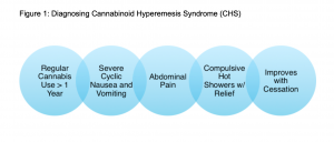
Cannabis is often used in cyclic vomiting syndrome (CVS) and chemotherapy-related nausea and vomiting due to its antiemetic properties. Paradoxically, hyperemesis as a result of chronic cannabis use has led to the recognition of a new disorder called cannabinoid hyperemesis syndrome (CHS). CHS first described in an Australian case series in 2004, is the syndromic clustering of nausea, cyclic vomiting and abdominal pain, often relieved with hot showers (emdocs) (2,3,4,6,11). Symptoms are often seen in chronic users with daily to near-daily exposure to cannabis. Many users mistakenly increase consumption assuming that intoxication will relieve symptoms. Thus far only total cannabis cessation has been found to be effective for elimination of symptoms (5,15,16).
Several theories exist about the cause of this syndrome, including pesticides on marijuana plants, however most believe it is due to the increased potency of THC with advancements in the cultivation of the marijuana plant. Some speculate that chronic high potency THC in genetically predisposed individuals causes differential degrees of cannabinoid receptor downregulation and autonomic dysregulation which can lead to nausea and vomiting (2, 5, 14, 24). Cannabis may have a biphasic mechanism of action, where at lower or less frequent doses it has anti‐emetic effects but at higher or more chronic doses it acts as a pro-emetic.
Cannabinoid receptors CB1 and CB2 are the main receptors responsible for THC’s effects on the body (3). Some authors postulate the cannabinoid receptors in the medulla allow for the antiemetic properties of THC, while the cannabinoid receptors in the gastrointestinal tract are suspected to be the source of symptoms due to dysregulation (4,14). Others believe the TRPV1 receptor (transient receptor potential vanilloid subtype 1), which is activated by marijuana, capsaicin and heat, is altered by chronic marijuana use, and responsible for CHS (8). Both endogenous and exogenous cannabinoids act as agonists for TRPV1.
Manny: While no diagnostic criteria currently exist for definitive CHS diagnosis, Sorenson et al performed a systematic review and identified six major characteristics patients typically display.
- History of regular cannabis use (100%)
- Cyclic nausea and vomiting (100%)
- Generalized, diffuse abdominal pain (85.1%)
- Compulsive hot showers with symptom improvement (92.3%)
- Symptoms resolve with marijuana use cessation (92.3%)
- A higher prevelance in males (72.9%)
Often patients will experience three phases of Cannabinoid Hyperemesis Syndrome (3,8):
- The Pre-emetic or Prodromal Phase:
- This phase can last for months or years, and is characterized by diffuse abdominal discomfort, feelings of agitation or stress, morning nausea, and fear of vomiting
- It may also include autonomic symptoms like flushing, sweating, and increased thirst
- Patients often have increased use of marijuana to treat these symptoms
- Hyperemetic Phase:
- This phase lasts 24-48 hours and is characterized by multiple episodes of vomiting with diffuse, severe abdominal pain
- Recovery Phase:
- This begins with total cessation of cannabis
- Often patients require a bowel regimen, IV fluids, and electrolyte replacement
- Resolution of symptoms may take up to one month
Brit: It is important to note that cannabinoid hyperemesis syndrome is a diagnosis of exclusion. If it is a patient’s first presentation for nausea, vomiting and abdominal pain, other primary etiologies such as gallbladder or intestinal disease, intoxications, or surgical emergencies should be considered, before a diagnosis of CHS is solidified. Patients with repeat presentations for nausea, vomiting and abdominal pain, with chronic marijuana use, negative previous evaluations and no history of diabetes, allow CHS to be higher on the differential diagnosis.
Most Common Complications of CHS:
- Electrolyte abnormalities (most commonly low potassium)
- Dehydration or acute kidney injury
- Muscle cramping or spasms
Documented Life-Threatening Complications of CHS:
- Pneumomediastinum from a ruptured esophagus
- Electrolyte derangement causing seizures, arrhythmias
Research on the treatment of CHS is limited. Clinicians often treat symptoms of CHS including nausea, vomiting, and abdominal pain with traditional pharmaceuticals. All patients with suspected CHS should be offered cannabis cessation counseling, resources, and follow up (15). Only total cannabis cessation has been found to be effective for preventing and eliminating symptoms (5,15,16).
Capsaicin
Capsaicin, the active ingredient of chili peppers, has been promoted as a treatment for CHS initially based on patients’ successes with hot showers. Several case series and retrospective reviews have shown benefits in adolescents (9) and adults (10). The cream is applied to the fatty areas of the backs of the arms and abdomen (4), and causes a sensation of warmth or burning. It comes in various concentrations between 0.025% to 0.15%, and is applied to the shoulders, extensor surface of the arms, back or abdomen up to three times daily. (Table 2.)
One proposed mechanism behind the relief of capsaicin is its ability to transiently activate TRPV1, previously down regulated during chronic exposure to cannabinoids. (8, 12). Capsaicin along with warm stimulation such as hot showers, activate TRPV1, which may explain the relief CHS patients receive from compulsive showering. Capsaicin is cheap, with few side effects, and is often successful for treating patients who have not received other medications for their CHS before (4).
Benzodiazepines
One or a combination of the following medications can be successful for acute treatment of CHS symptoms. A systematic review of pharmacological treatments for CHS found evidence to support benzodiazepines such as lorazepam and diazepam (20). These medications work through GABA receptor agonism, thus neurotransmitter inhibition, causing sedation, anxiolysis, and muscle relaxation.
Antipsychotics and Tricyclic Antidepressants
Previously reserved for agitated patients, haloperidol has been used off-label for postoperative nausea and has been often used successfully to treat CHS. Several case reports describe complete relief of symptoms from haloperidol (18,19). The drug is a butyrophenone antipsychotic with high affinity dopamine antagonism at the D2 receptor in the CNS. Currently a randomized crossover clinical trial is completing research comparing haloperidol and ondansetron (21). Likely not a medication to start in the emergency department, tricyclic antidepressants have demonstrated effectiveness in cyclic vomiting syndrome and CHS (20,21). It is believed to be related to serotonin reuptake inhibition in addition to antihistamine effects (23). Unfortunately the above medications are not without risk.While capsaicin adverse reactions are topical and non-life-threatening, some of the listed medications can cause significant side-effects including Qt prolongation and arrhythmias, respiratory depression, and extreme feelings of anxiety.
Future Research
We know emergency departments are witnessing increasing incidents of cannabis related pathology (1). A significant group of these patients suffer from CHS, and many remain undiagnosed. These patients often receive expansive diagnostic workups, numerous pharmacological interventions, and frequently require observation or hospitalization. A recent systematic review found poor quality of evidence beyond case reports many pharmaceuticals (20) and no randomized trials have been completed. While research continues to be in its early stages, the early recognition of CHS and treatment may prevent costly work ups, admissions and prolonged symptoms of patients suffering from CHS (17).
Manny: That rounds out our summary of the key emDocs posts. Thanks for joining us on the podcast, and stay tuned for our next episode. Feel free to comment on our site and let us know if you have any feedback. Stay safe and healthy everyone!